Quantcast
Channel: New Drug Approvals
Viewing all 2881 articles
Browse latest View live

Are You Efficient? Why R&D Process Streamlining Could Affect 140,000 Jobs

$
0
0

Originally posted on AAPS Blog:

By Robert G. Bell

Robert BellA recent survey and report by PhRMA and Battelle indicates that pharmaceutical executives believe there will be strong growth in R&D and manufacturing but that fewer workers would be needed as “efficiencies” in R&D and manufacturing take hold in the next 10 years.

Pharmaceutical executives were surveyed for their best-case, most likely case, and worst-case views and projections for the future growth probabilities of U.S. and global pharmaceutical industry across R&D and manufacturing activities for the next 10 years. These different potential futures for domestic growth in pharmaceutical R&D and manufacturing activities are dependent on policy choices made in the United States regarding coverage/payment to encourage innovation, regularity systems, and intellectual property. The report’s authors developed a statistically adjusted or normalized distribution of future growth probabilities based on the distribution of these future growth rates and how much, on average, the individual scores varied from the current…

View original 404 more words


Filed under: Uncategorized

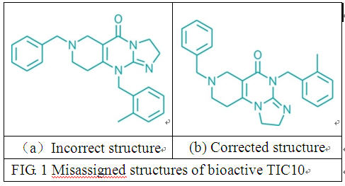
TIC 10 structure established……Synthesis: Structural misassignment stems from long-standing use of incorrect recipe to prepare anticancer agent

$
0
0

09221-notw1-strucs

Misassigned (top) and corrected (bottom) structures of bioactive TIC10.


PIC FROM

http://pipeline.corante.com/archives/2014/05/22/a_horrible_expensive_and_completely_avoidable_drug_development_mixup.php

Don’t forget the chemistry
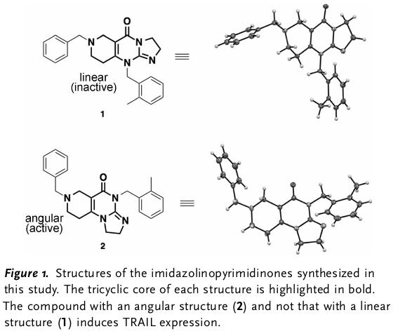
In a bit of a whoopsie it has come to light 1 that a compound TIC10, a stimulator of gene expression for TRAIL and in PhI/II clinical trials has in fact got an incorrect structure or rather the compound that was protected in the patent was assigned the incorrect structure. It was patented in a single compound patent 2 – which might ring alarm bells for some – were they really that confident they had the best compound. A quick look at the patent revealed large amounts of in vitro work but none in vivo again begging the question how good is this single compound. The compound had been identified by a group at Pennsylvania State University and licensed to Oncoceutics from screening of the NCI compound collection but the team, as reported, only attempted structural characterisation by MS. This would be unlikely to differentiate regioisomers which is what the problem turns out to be. It does however seem strange that the patenting error was not detected during resynthesis and scale-up for progression to the clinic. The error was picked up when a group from the Scripp’s 3 who synthesised the patented compound but found it inactive while they found the NCI batch to be active. They characterised the patented (inactive) and non-patented (active ex NCI) structures by crystallography and total synthesis. The corrected structure has now been patented by the Scripps group and licensed to Sorrento.
Of course this is all a bit embarrassing for those concerned but also more seriously could end up with extensive patent litigation, wasting money and discouraging investors from supporting the work until the patent situation is clarified causing delay in progressing the asset. Please please talk to medicinal chemsist early in a project this one looks like no one did which has led to an expensive mistake.


1. S. Borman Chem Eng News 2014, May 26 page 7
2. US Patent US8673923
3. N. T. Jacob et al Angew. Chemie. Int. Ed., Article first published online: 18 May 2014 DOI: 10.1002/anie.201402133

TIC10-2
BOEHRINGER RECIPE
This synthesis from simpler starting materials, taken from the Boehringer 1973 patent, makes bioactive TIC10 with a three-fused-ring angular structure (tint), not the linear structure shown in the reaction scheme below and in the patent.

The fog is beginning to lift on how a mistake in the structural analysis of a promising drug candidate occurred and was sustained for so long that the agent was nearly in human clinical trials before the error was discovered.

Kim D. Janda and coworkers at Scripps Research Institute California recently discovered that the structure of a promising cancer drug candidate, called TIC10 or ONC201, had been misassigned in the agent’s patent (Angew. Chem. Int. Ed. 2014, DOI: 10.1002/anie.201402133). The biotech firm Oncoceutics, which has licensed the patent (U.S. 8673923), is sponsoring Phase I/II human clinical trials for the agent, which are currently in a prerecruitment phase. But Scripps has applied for a patent on the corrected structure and has licensed it exclusively to another company, Sorrento Therapeutics (C&EN, May 26, page 7).

TIC10 originated with a 1973 German patent (2150062) owned by C. H. Boehringer Sohn, in Ingelheim, now called Boehringer Ingelheim. The now-expired patent covers a family of 43 compounds, one of which is now called TIC10, and their possible use as antiseizure medications. It also contains a recipe for synthesizing the compounds.

At some point, the National Cancer Institute (NCI) picked up TIC10 for its publicly accessible Diversity Set II database, which researchers can screen freely to find agents with interesting activities. The compound’s structure shown in the database listing was the same as in the German patent—with three rings fused in a linear fashion.

Wafik S. El-Deiry of Pennsylvania State University and coworkers discovered that NCI’s TIC10 sample had potent anticancer activity (Sci. Transl. Med. 2013, DOI: 10.1126/scitranslmed.3004828). They used mass spectrometry to try to confirm that the structure of the compound was the same as that listed in the NCI database. But MS is inadequate for structure confirmation when used on its own.

Penn State was granted a patent to use TIC10 to treat cancer, and it licensed the patent to Oncoceutics for development. The company used the Boehringer recipe to develop a production process to produce sufficient amounts of the compound for study and eventually for clinical trials.

When Janda’s group decided to study TIC10 for possible use in a combination therapy, they opted to synthesize the three-ring linear structure from scratch, instead of using the Boehringer recipe. The compound they made that had that structure was bioinactive, so they ordered the compound from NCI and found that agent to be bioactive. When they then analyzed the bioactive agent carefully, they found it to have a structure in which one of the three fused rings is at an angle to the other two. They then synthesized this angular structure from scratch, applied for a patent on it, and relicensed it.

The reason the structural problem with TIC10 persisted so long is that until Janda’s group joined the TIC10 game, others all seemed to be using the Boehringer recipe to make it and were thus synthesizing a bioactive but structurally misidentified compound.

Oncoceutics Chief Business Officer Lee Schalop notes that all of the company’s research on ONC201, including studies required for approval of the agent’s Investigational New Drug Application with the Food & Drug Administration, were carried out with the bioactive agent. Oncoceutics did not reply by C&EN press time to an inquiry about what techniques it used to characterize ONC201 during its studies.

A number of reagent suppliers have also been marketing TIC10 for research purposes. A search of Chemical Abstracts Service databases in May showed that eight companies were selling CAS Registry Number 41276-02-2, the compound with the misassigned linear structure. (CAS is a unit of the American Chemical Society, which publishes C&EN.)

Sigma-Aldrich, one of the companies offering TIC10, hired a contractor to synthesize TIC10. But the company didn’t officially launch the compound for sale and didn’t ship any to customers. In response to the Janda publication, Sigma-Aldrich halted the product launch.

Sorrento patent attorney Jeff Oster tells C&EN that a meeting between Oncoceutics and Sorrento had been scheduled to begin untangling some of TIC10’s patent and licensing issues. But Schalop says Oncoceutics has no plans to meet with Sorrento.


Filed under: Uncategorized Tagged: TIC 10

Immunomedics’ IMMU-132 Gets Orphan Drug Status For Small Cell Lung Cancer

$
0
0


(RTTNews) – Immunomedics, Inc. (IMMU), a biopharmaceutical company focusing mainly on the development of monoclonal antibody-based products for the targeted treatment of cancer, autoimmune and other serious diseases, said its antibody-drug conjugate for solid cancer therapy, IMMU-132, has received orphan drug status from the Office of Orphan Products Development of the U.S. Food and Drug Administration or FDA for small cell lung cancer or SCLC treatment.

http://www.quotenet.com/news/stocks/Immunomedics-IMMU-132-Gets-Orphan-Drug-Status-For-Small-Cell-Lung-Cancer-782707

Immunomedics has received orphan drug designation from the US Food and Drug Administration’s (FDA) Office of Orphan Products Development for its IMMU-132 for pancreatic cancer therapy.

IMMU-132 is Immunomedics’ antibody-drug conjugate in clinical development for treatment of patients with solid cancer.

Immunomedics president and CEO Cynthia Sullivan said that this is the second orphan designation from FDA for IMMU-132, which has demonstrated activity in patients with advanced pancreatic cancer, as well as partial responses in five other types of solid cancer.

“The humanised antibody internalises into cancer cells following binding to TROP-2, making it a suitable candidate for the delivery of cytotoxic drugs.”

The FDA previously granted orphan drug designation to IMMU-132 for treatment of small-cell lung cancer patients.

In an ongoing Phase I/II clinical study, IMMU-132 has resulted in partial responses in patients with colorectal cancer, esophageal cancer, triple negative breast cancer, and small-cell and non-small-cell lung cancers.

IMMU-132 is composed of a humanised antibody, hRS7, that binds to the trophoblast cell-surface antigen (TROP-2), also known as the epithelial glycoprotein-1 antigen (EGP-1).

The humanised antibody internalises into cancer cells following binding to TROP-2, making it a suitable candidate for the delivery of cytotoxic drugs.

In preclinical studies, IMMU-132 has demonstrated that it delivers 120-times the amount of SN-38, the active metabolite of irinotecan, to a human pancreatic tumor xenograft than when irinotecan is given.

IMMU-132 significantly improves survival and tumour regression in various animal models of human cancers.


Filed under: 0rphan drug status Tagged: IMMU-132, Immunomedics, Orphan Drug Status, Small Cell Lung Cancer

EffRx Pharmaceuticals receives FDA orphan drug designation for EX404

$
0
0


EffRx Pharmaceuticals has received US Food and Drug Administration (FDA) orphan-drug designation for its proprietary metformin-based product, EX404, for treatment of paediatric polycystic ovary syndrome (PCOS).

Also known as Stein-Leventhal syndrome, PCOS is a heterogeneous disorder of chronic anovulation and hyperandrogenism.

The syndrome is believed to occur due to hormonal imbalance caused by increased levels of androgens and insulin in the body.

EffRx Pharmaceuticals chairman and CEO Christer Rosén said the FDA’s orphan drug designation of EX404 is a significant step forward in the clinical development programme.

http://www.pharmaceutical-technology.com/news/newseffrx-pharmaceuticals-receives-fda-orphan-drug-designation-for-ex404-4290237?WT.mc_id=DN_News


Filed under: 0rphan drug status Tagged: EffRx Pharmaceuticals, EX404, fda, Orphan Drug Designation

Momenta Pharma receives FDA orphan drug designation for pancreatic cancer drug Necuparanib

$
0
0

heparan sulfate mimetic derived from unfractionated heparin with a molecular weight between 5500 and 6200 Da


Necuparanib

M-402
M-ONC-402
MONC 402


Momenta Pharmaceuticals Inc

http://www.pharmaceutical-technology.com/news/newsmomenta-pharma-receives-fda-orphan-drug-designation-pancreatic-cancer-drug-4287892

Momenta Pharmaceuticals has received orphan drug designation from the US Food and Drug Administration (FDA) for its necuparanib, a heparan sulfate mimetic indicated for treatment of pancreatic cancer.

Momenta Pharmaceuticals chief medical officer Jim Roach said there is a great need for new medications for patients suffering from pancreatic cancer.

“We are encouraged by the progress of the programme to date, and in the next several months, we anticipate completing Part A of our ongoing Phase I/II study of necuparanib in combination with Abraxane and gemcitabine,” Roach said.

“In the next several months, we anticipate completing Part A of our ongoing Phase I/II study of necuparanib in combination with Abraxane and gemcitabine.”

“We look forward to sharing the results from Part A and advancing the product into the Phase II part of the study in the second half of 2014.”

Necuparanib has recently been adopted as the unique non-proprietary name for M402 by The United States Adopted Names.

The drug is derived from unfractionated heparin. It has been engineered to have significantly reduced anticoagulant activity while preserving the relevant antitumor properties of heparin.

Part A dose escalation component of the Phase I/II trial, which is evaluating necuparanib in combination with Abraxane (nab-paclitaxel) and gemcitabine in advanced metastatic pancreatic cancer patients, is expected to be completed in the next several months.

The company is expected to report the clinical data from Part A in the second half this year. The company also plans to begin Part B of the study by the year-end.

Part B will be a randomised, controlled, proof-of-concept study to assess the antitumor activity of necuparanib in combination with Abraxane plus gemcitabine, versus Abraxane plus gemcitabine alone.


Heparin, a highly sulfated heparin-like glycosaniinoglycan (HLGAG) produced by mast cells and isolated from natural sources, is a widely used clinical anticoagulant. However, the effects of natural, or unfractionated, heparin can be difficult to predict and patients must be monitored closely to prevent over- or under-anticoagulation. Low molecular weight heparins (LMWHs) obtained by various methods of fractionation or depolymerization of polymeric heparin have more predictable pharmacological action as anticoagulants, reduced side effects, sustained antithrombotic activity, and better bioavailability than unfractionated heparin (UFH). Several LMWHs are approved for outpatient treatment of thrombotic conditions.

There is increasing interest in the potential role of antithrombotic agents in the management of cancer patients. Results from several recent clinical trials have suggested a survival advantage for certain types of cancer patients treated with LMWHs (reviewed in Lemoine, 2005, Journal of Clinical Oncology, 23: 2119-20).


http://www.google.fm/patents/EP2207811A1?cl=en

The invention is based, in part, on the development of polysaccharide preparations, e.g., preparations of polysaccharides derived from heparin, that lack substantial anticoagulant activity (e.g., preparations of polysaccharides that have substantially no anticoagulant activity) but retain activity in other non-coagulation mediated biological processes, and methods to produce them. These compounds can have one or more of the following features: 1) an anti-Xa activity and an anti-IIa activity each less than 50 IU/mg, and 2) anti-metastatic, anti-angiogenic, anti-fibrotic and/or anti-inflammatory activity. The polysaccharides disclosed herein can also have structural characteristics that distinguish them from other polysaccharides, (e.g., from commercially available heparins). For example, a polysaccharide preparation provided herein can have one or more of the following characteristics: the preparation has less than 50% glycol split uronic acid residues; the preparation has no more than 3 glycol split uronic acid residues (UG) per polysaccharide chain; the preparation has greater than 40% U2SHNS>6S disaccharide residues; degree of desulfation of the preparation is less than 40%; one or more polysaccharide chains in the preparation have a 4,5-unsaturation of a non-reducing end uronic acid residue; one or more polysaccharide chains in the preparation have a 2,5-anhydromannitol residue at the reducing end; and the weight average molecular weight of the preparation is between 3,500 and 7,000 Da. This disclosure includes preparations having one or more of these properties and characteristics as well as methods of making and using such preparations. The disclosure also features methods of using such preparations.

Accordingly, in a first aspect, the invention features a polysaccharide preparation (e.g., a heparin-derived preparation) having the following characteristics: (a) a weight average chain molecular weight between 3,500 and 7,000 Da; (b) an anti-Xa activity and an anti-IIa activity each less than 50 IU/mg (e.g., an anti-Xa activity less than about 40 IU/mg, 30 IU/mg, 20 IU/mg, 15 IU/mg, or 10 IU/mg and an anti-IIa activity less than about 40 IU/mg, 30 IU/mg, 20 IU/mg, 10 IU/mg, 5 IU/mg, 4 IU/mg, or 3 IU/mg); and (c) less than 50% glycol split uronic acid residues (e.g., less than 40%, 30%, 25%, or 20% glycol split uronic acid residues) in the preparation. In some embodiments, the preparation contains between 5% and 50% glycol split uronic acid residues (e.g., between 5% and 40%, 5% and 30%, 10% and 50%, 10% and 40%, or 10% and 30% glycol split uronic acid residues).

In a second aspect, the invention features a polysaccharide preparation (e.g., a heparin- derived preparation) having the following characteristics: (a) a weight average chain molecular weight between 3,500 and 7,000 Da; (b) an anti-Xa activity and an anti-IIa activity each less than 50 IU/mg (e.g., an anti-Xa activity less than about 40 IU/mg, 30 IU/mg, 20 IU/mg, 15 IU/mg, or 10 IU/mg and an anti-IIa activity less than about 40 IU/mg, 30 IU/mg, 20 IU/mg, 10 IU/mg, 5 IU/mg, 4 IU/mg, or 3 IU/mg); and (c) the polysaccharide chains of the preparation have no more than 3 glycol split uronic acid residues (UQ) per polysaccharide chain (e.g., each polysaccharide chain has no more than 2 or no more than 1 glycol split uronic acid residue (UQ) per polysaccharide chain).

In a third aspect, the invention features a polysaccharide preparation (e.g., a heparin- derived preparation) having the following characteristics: (a) a weight average chain molecular weight between 3,500 and 7,000 Da; (b) an anti-Xa activity and an anti-IIa activity each less than 50 IU/mg (e.g., an anti-Xa activity less than about 40 IU/mg, 30 IU/mg, 20 IU/mg, 15 IU/mg, or 10 IU/mg and an anti-IIa activity less than about 40 IU/mg, 30 IU/mg, 20 IU/mg, 10 IU/mg, 5 IU/mg, 4 IU/mg, or 3 IU/mg); and (c) polysaccharide chains of the preparation have on average no more than 3 glycol split uronic acid residues (Uo) per polysaccharide chain (e.g., on average no more than 2.5, no more than 2, no more than 1.5, or no more than 1 glycol split uronic acid residues (UG) per polysaccharide chain.

In a fourth aspect, the invention features a polysaccharide preparation (e.g., a heparin- derived preparation) having the following characteristics: (a) a weight average chain molecular weight between 3,500 and 7,000 Da; (b) an anti-Xa activity and an anti-IIa activity each less than 50 IU/mg (e.g., an anti-Xa activity less than about 40 IU/mg, 30 IU/mg, 20 IU/mg, 15 IU/mg, or 10 IU/mg and an anti-IIa activity less than about 40 IU/mg, 30 IU/mg, 20 IU/mg, 10 IU/mg, 5 IU/mg, 4 IU/mg, or 3 IU/mg); and (c) the preparation has greater than 40% U2SHNS,6S disaccharide residues (e.g., greater than 50%, 60%, 70%, or 80% U2SHNS,6S disaccharide residues). In some embodiments, the preparation has a degree of desulfation less than 40% (e.g., less than 30%, 20%, or 10%).

In a fifth aspect, the invention features a polysaccharide preparation (e.g., a heparin- derived preparation) lacking substantial anticoagulant activity (e.g., having substantially no anticoagulant activity), wherein the preparatiorrmdudes-polv^accharides that include Formula I:

[Uw-HXjy)Z]m~[UG-HX5y5Z]n

wherein U indicates a uronic acid residue and H indicates a hexosamine residue; m and n are integers such that m = 4-16 (e.g., 4-8, 4-9, 4-10, 4-11, 4-12, 4-13, 4-14, or 4-15), and n = 1-4 (e.g., 1-2 or 1-3);

w = -2OS or -2OH; x = -NS or -NAc; y = -3OS or -3OH; z = -60S or -6OH;

wherein the symbol ~ indicates that the units marked m and n are distributed along the polysaccharide chain and are not necessarily in sequence, wherein w, x, y, and z are each the same or different on each unit marked m, and wherein x, y, and z are each the same or different on each unit marked n.

In a sixth aspect, the invention features a polysaccharide preparation (e.g., a heparin- derived preparation) lacking substantial anticoagulant activity (e.g., having substantially noanticoagulant activity) and having antimetastatic activity, wherein the preparation includes polysaccharides that include Formula II:

[Uw-HXjy;Z] m- [UG-HX)y;Z] n- [Uw-HX;y)Z] 0- [UG-HX^2] p- [Uw-HX;yjZ] q

wherein U indicates a uronic acid residue and H indicates a hexosamine residue; wherein m-r are integers such that: m = 0-10; n= 0- 3;

O = O-IO;

P = 0-3; q = 0-10;

w = -2OS or -2OH; x = -NS or -NAc; y = -3OS or -3OH; z = -60S or -6OH;

wherein w, x, y, and z are each the same or different on each unit marked m, n, o, p, or q. In some embodiments, the sum of n + p is less than or equal to 4 (e.g., less than or equal to 3, 2, 1, or 0). In some embodiments, the preparation has a weight average chain molecular weight between 3,500 and 7,000 Da.


Examples of such polysaccharide preparations include chains that include the following:

[Uw-HX;yjZ]m~[UG-Hx y z]n

wherein U indicates a uronic acid residue and H indicates a hexosamine residue, wherein m and n are integers such that m = 6-18, and n = 1 -4, w = -2OS or -2OH, x = -NS or -NAc, y = -3OS or -3OH, z = -60S or -6OH,


wherein the symbol ~ indicates that the units marked m and n are distributed along the polysaccharide chain and are not necessarily in sequence, wherein w, x, y, and z are each the same or different on each unit marked m, and wherein x, y, and z are each the same or different on each unit marked n; and

[Uw-HX)y)Z]m-[UG-HXiy)Z]n-[Uw-HXjyjZ]o-[UG-HX5y)Z]p-[Uw-HX!yiZ]q

wherein U indicates a uronic acid residue and H indicates a hexosamine residue, wherein m-r are integers such that: m = 0-10, n= 0- 3, o = 0-10, p = 0-3, q = 0-10, w = -2OS or -2OH, x = -NS or -NAc, y = -3OS or -3OH, z = -60S or -6OH,

wherein w, x, y, and z are each the same or different on each unit marked m, n, o, p, or q.

Anti-IIa Activity

Polysaccharide preparations are disclosed herein that provide substantially reduced anti- Ha activity, e.g., anti-IIa activity of about 0 to 50 IU/mg, about 0 to 40 IU/mg, about 0 to 30 IU/mg, about 0 to 25 IU/mg, about 0 to 20 IU/mg, about 0 to 10 IU/mg, about 0 to 5 IU/mg, about 5 to 10 IU/mg, about 5 to 15 IU/mg, about 5 to 20 IU/mg. Anti-IIa activity is calculated in International Units of anti- Ha activity per milligram using statistical methods for parallel line assays. The anti-IIa activity levels described herein are measured using the following principle.

Polysaccharide (PS) + ATIII→ [PS • ATIII]

Ha

PS • ATIII→[PS • ATIII • Ha] + Ha (Excess)

Ha (Excess) + Substrate -» Peptide + pNA (measured spectrophotometrically) Anti-factor Ha activity is determined by the sample potentiating effect on antithrombin (ATIII) in the inhibition of thrombin. Thrombin excess can be indirectly spectrophotometrically measured. The anti-factor Ha activity can be measured, e.g., on a Diagnostica Stago analyzer or on an ACL Futura3 Coagulation system, with reagents from Chromogenix (S-2238 substrate, Thrombin (53 nkat/vial), and Antithrombin), or on any equivalent system. Analyzer response is calibrated using the 2nd International Standard for Low Molecular Weight Heparin.


Filed under: 0rphan drug status, Phase2 drugs Tagged: M 402, Necuparanib

Grape-enriched diet supports eye health

$
0
0

Originally posted on lyranara.me:

New research presented this week at the Association for Research in Vision and Ophthalmology conference in Orlando, Florida suggests that regular grape consumption may play a role in eye health by protecting the retina from deterioration. Specifically, a grape-enriched diet resulted in a protective effect on retinal structure and function.

The retina is the part of the eye that contains the cells that respond to light, known as photoreceptors. There are two types of photoreceptors: rods and cones. Retinal degenerative diseases affect over 5 million people in the U.S., and can cause blindness due to photoreceptor cell death.

The study was conducted by a research team at the University of Miami, Bascom Palmer Eye Institute and investigated whether a diet supplemented with grapes could protect the photoreceptors in mice with retinal degeneration. Mice were either fed a grape-supplemented diet corresponding to 3 servings of grapes per day for humans…

View original 136 more words


Filed under: Uncategorized

Grape consumption may offer benefits for symptomatic knee osteoarthritis

$
0
0

Originally posted on lyranara.me:

New research presented last week at the Experimental Biology conference in San Diego, California, suggests that regular grape consumption may help alleviate pain associated with symptomatic osteoarthritis of the knee, and improve joint flexibility and overall mobility. Researchers attribute these potential benefits to the polyphenols found in grapes.

The sixteen week clinical study, undertaken by Texas Woman’s University, was designed to investigate the benefits of grape consumption on inflammation and osteoarthritis outcomes. 72 men and women with knee osteoarthritis (OA) were assigned to either consume grapes in the form of a whole grape freeze-dried powder, or a placebo powder.

The study results, presented by lead investigator Shanil Juma, Ph.D., showed that both men and women consuming a grape-enriched diet had a significant decrease in self-reported pain related to activity and an overall decrease in total knee symptoms. This beneficial effect was more pronounced in females. Additionally, age-related differences were observed:…

View original 374 more words


Filed under: Uncategorized

Latin American Active Pharmaceutical Ingredients Industry Catches Up on the US

$
0
0
South and Central America's active pharmaceutical ingredients market are about to close the gap on the northern neighbors...
South and Central America’s active pharmaceutical ingredients market are about to close the gap on the northern neighbors… (Picture: PROCESS India)

Latin American Active Pharmaceutical Ingredients Industry Catches Up on the US

The market for active pharmaceutical ingredient in the Americas shows a clear north–south divide: 88% percent for the US and Canada, the rest for South and Central American companies. but as the economy in Latin America booms and prosperity growths, these markets are just about to catch up on the US new figures indicate….http://www.process-worldwide.com/management/markets_industries/articles/374702/


Filed under: PROCESS Tagged: API, PROCESS

Design, construction and start-up of a pilot plant for active pharmaceutical ingredients

$
0
0
Linde-KCA-Dresden built a pilot plant 16 times the size of the laboratory apparatus for Novo Nordisk A/S
The building layouts show different areas for the pilot plant, the utility systems and the technical rooms with hazardous and non-hazardous proof conditions (top). The engineering documents include the piping and instrumentation diagrams, the manufacturer vessel drawings as well as the 3D model with the isometric drawings of the pipes (bottom)
The building layouts show different areas for the pilot plant, the utility systems and the technical rooms with hazardous and non-hazardous proof conditions (top). The engineering documents include

The construction of a pilot plant constitutes an important milestone in the development cycle of a new active pharmaceutical ingredient. Pilot plants are used to gain the technological experience needed for the scale-up process and are required for producing sufficient quantities of the active pharmaceutical ingredient for clinical trials and other types of tests. As a result, even these testing facilities must comply with GMP regulations.

http://www.cpp-net.com/pharma/-/article/5829537/15910929/Challenging+task/


Filed under: Drug discovery Tagged: API, drugs, PROCESS

Polyphenols and Alzheimers

$
0
0

Originally posted on Ayurvedic Herbal Teas:

“Alzheimer’s disease (AD) is the most prevalent neurodegenerative disease in the growing population of elderly people. A hallmark of AD is the accumulation of plaques in the brain of AD patients. The plaques predominantly consist of aggregates of amyloid-beta (Abeta), a peptide of 39-42 amino acids generated in vivo by specific, proteolytic cleavage of the amyloid precursor protein.” (Finder & Glockshuber)

Stefani & Rigacci review the evidence for polyphenols and their ability to reduce amyloid aggregation. Natural polyphenols are emerging as an increasingly attractive treatment for amyloid disease prevention and therapy.  Evidence suggests that they can inhibit the production of amyloidogenic peptides, increase antioxidant enzyme activity and reduce inflammation. The researchers suggest that we should now be describing them as potentially multitargeting drugs.

Potential sources of polyphenols are found in, amongst others, vegetables, fruit and green tea.

YOUNGFUN_2013_01

Green Tea Black Bacground

View original


Filed under: Uncategorized

cGMPs for Active Pharmaceutical Ingredient (API) and Excipient Production

$
0
0

Originally posted on DRUG REGULATORY AFFAIRS INTERNATIONAL:

For many years the only Food and Drug Administration
(FDA) document specifically related to current good
manufacturing practice (cGMP) requirements for active
pharmaceutical ingredient (API) manufacturers was a guid-
ance for FDA investigators that covered all Bulk
Pharmaceutical Chemicals (BPCs). Over the past few years,
two iterations of a DRAFT guidance for industry specifically
addressing API manufacture were published. On September
25, 2001, the lack of specific, official cGMP guidance for
API manufacturers came to an end when “FDA’s Guidance
for Industry:
Q7A Good Manufacturing Practice Guidance
for Active Pharmaceutical Ingredients (“Q7A”)”
read at

View original


Filed under: Uncategorized

BI-836845 a fully human mAb targeting IGF-1 created using HuCAL technology from Morphosys, for the potential iv infusion treatment of cancer, including solid tumors and breast cancer.

$
0
0

BI-836845

Human monoclonal IgG1 lambda antibody against IGF-1 (insulin growth factor-1) and IGF-2

IGF pathway modulator (iv, cancer),  Boehringer Ingelheim;

Phase 2 Clinical

Anticancer protein kinase inhibitor; Anticancer monoclonal antibody

WO-2008155387

Boehringer Ingelheim International Gmbh

Boehringer Ingelheim is developing BI-836845, a fully human mAb targeting IGF-1 created using HuCAL technology from Morphosys, for the potential iv infusion treatment of cancer, including solid tumors and breast cancer.

In April 2011, a phase I trial was initiated in the UK . In October 2011, another phase I trial was initiated in Taiwan. In February 2014, recruitment was ongoing. At that time, the trial was expected to be completed in March 2015 In June 2014, the drug was listed as being in phase I development for solid tumors in Japan and for breast cancer

In May 2014, an open-label, randomized, parallel-assigned, phase II trial (NCT02123823; 1280.4; 2013-001110-15) to evaluate the safety and efficacy of BI-836845 and everolimus in combination with exemestane in women with breast cancer (expected n = 198) was planned to be initiated in Belgium, France and the Netherlands. At that time, the trial was expected to complete in December 2017

In June 2014, an open-label, single-group assigned, phase I trial (NCT02145741; 1280.15) to evaluate BI-836845 in Japanese patients (expected n = 18) with advanced solid tumors was planned to be initiated in Japan. At that time, the trial was expected to complete in June 2015
In March 2011, a non-randomized, open-label, phase I study (NCT01317420; 1280.2; 2010-021714-29) was planned to begin later that month in patients with solid tumors (expected n = 70) in the UK, to assess the safety, efficacy, pharmacokinetics, pharmacodynamics and pharmacogenomics of BI-836845. The study began in April 2011; at that time, completion was expected in March 2013 .

In June 2012, preclinical data were presented at the 48th ASCO meeting in Chicago, IL. In the study, the combination of BI-836845 plus rapamycin was more effective than single agent therapy at inhibiting Ewing’s sarcoma cell proliferation in vitro and in a nude mouse xenograft model .

In November 2011, preclinical data were presented at the 23rd AACR-NCI-EORTC International Conference in San Francisco, CA. BI-836845 potently inhibited proliferation of the multiple myeloma cell line LP-1 with an EC50 of 0.4 nM.

BI-836845 is a human monoclonal IgG1 lambda antibody against IGF-1 (insulin growth factor-1) and IGF-2 (insulin growth factor-2). Phase II clinical trials are ongoing at Boehringer Ingelheim for the treatment of patients with breast cancer, and phase I clinical trials are ongoing with patients with advanced solid tumors.

Insulin-like growth factor-1 (IGF-1; a 70 amino-acid polypeptide) and insulin-like growth factor-2 (IGF-2; a 67 amino-acid polypeptide) are 7.5-kD soluble factors present in serum that can potently stimulate the growth of many mammalian cells (reviewed by Pollack et al., 2004). Although IGFs can be detectable in a number of tissues the main source of circulating IGFs is the liver which secretes the IGFs and IGF binding proteins (IGFBPs) in response to a complex signaling pathway that is initiated in the pituitary gland and transduced via growth hormone. On secretion into the bloodstream the IGFs form complexes with the IGFBPs which not only protects them from proteolytic degradation in the serum en route to their target tissues but also prevents their association with the IGF receptors. In addition to this endocrine source of IGFs they are also known to be secreted in an autocrine or paracrine manner in target tissues themselves. This is known to occur during normal fetal development where the IGFs play a key role in the growth of tissues, bone and organs. It is also seen in many cancer tissues where there is thought to be paracrine signaling between tumour cells and stromal cells or autocrine IGF production by the tumour cells themselves (reviewed by LeRoith D, 2003).

30 May 2014

MEDIA ALERT

ASCO 2014: Boehringer Ingelheim to present latest oncology research, including overall survival results

• Highly anticipated new overall survival data for Giotrif® (afatinib*) to be presented on June 2nd (3:00 – 6:00 PM, E Hall D2 [Abstract #8004 scheduled for 4:00 - 4:12 PM])
• 7 total abstracts accepted for Giotrif® (afatinib*), nintedanib** and BI 836845**: 1 for oral presentation and 6 posters

BI 836845 (IGF ligand antibody)**
A Phase I dose escalation study of weekly BI 836845, a fully human, affinity-optimized, insulin-like growth factor (IGF) ligand neutralizing antibody, in patients with advanced solid cancers Chia-Chi Lin, Kwang-Yu Chang, Dennis Chin-Lun Huang, Vicky Marriott, Ludy van Beijsterveldt, Li-Tzong Chen, Ann-Lii Cheng Sunday, June 1
8:00 – 11:45 AM
S Hall A2
(Abstract #2617
Poster #80)
Phase I dose escalation study of 3-weekly BI 836845, a fully human, affinity optimized, insulin-like growth factor (IGF) ligand neutralizing antibody, in patients with advanced solid tumours Rihawi K, Ong M, Michalarea V, Bent L, Buschke S4, Bogenrieder T, Anthoney A, de Bono J, Twelves CJ Sunday, June 1
8:00 – 11:45 AM
S Hall A2
(Abstract #2622
Poster #85)


The activity of the IGFs is thought to be regulated by a complex and relatively poorly understood interaction involving seven different IGFBPs and other serum proteins. Activation of the IGFs involves their release from this ternary complex after proteolytic release of the serum binding protein and IGFBPs, this is thought to occur in close proximity to cell surfaces where the IGFs are then free to bind to their receptors and transduce intracellular signals that ultimately leads to cellular proliferation and the inhibition of apoptosis. IGF-1 and IGF-2 are able to bind to the IGF-1 receptor (IGF-1R) expressed on many normal tissues, which functionally is a 460 kD heterotetramer consisting of a dimerised alpha- and beta-subunit, with similar affinities (Rubin et al., 1995). IGF-2 can also bind to the IGF-2 receptor (also know as the mannose-6-phosphate receptor) which does not have any known signaling function, rather it is thought to act as a sink for IGF-2 and prevent it from binding and signaling through the IGF-1R. In this respect the IGF-2R has been demonstrated to be a tumour suppressor protein. The IGF-1R is structurally similar to the insulin receptor which exists in two forms, IR-A and IR-B, which differ by an alternatively spliced 12 amino acid exon deletion in the extracellular domain of IR-A. IR-B is the predominant IR isoform expressed in most normal adult tissues where it acts to mediate the effects of insulin on metabolism. IR-A on the other hand is known to be highly expressed in developing fetal tissues but not in adult normal tissues. Recent studies have also shown that IR-A, but not IR-B, is highly expressed in some cancers. The exon deletion in IR-A has no impact on insulin binding but does cause a small conformational change that allows IGF-2 to bind with much higher affinity than for IR-B (Frasca et al., 1999; Pandini et al., 2002). Thus, because of it’s expression in cancer tissues and increase propensity for IGF-2 binding, IR-A may be as important as IGF1-R in mediating the mitogenic effects of IGF-2 in cancer.

Binding of the IGFs to IGF-1R triggers a complex intracellular signaling cascade which results in activation of proteins that stimulate growth and inhibit apoptosis (reviewed by Pollack et al., 2004). In terms of growth, upregulated translation is induced by the activation of p70 S6 kinase, which in turn phosphorylates the S6 ribosomal protein (Dufner and Thomas, 1999). Thus, IGF-stimulated cell growth can be measured by the rapid increase in phosphorylated S6 ribosomal protein.

Unlike the EGFR and Her2neu receptors there is no known amplification of the IGF1-R or IR-A receptors in cancers indicating that receptor activation is controlled by the presence of active ligand. There is a very large body of scientific, epidemiological and clinical literature implicating a role for the IGFs in the development, progression and metastasis of many different cancer types (reviewed by Jerome et al., 2003; and Pollack et al., 2004).

For example, in colorectal cancer the expression of IGF-2 mRNA and protein is elevated in clinical colorectal tumour specimens compared with adjacent normal tissue (Freier et al., 1999; Li et al., 2004). There is also a positive correlation of elevated IGF serum levels with proliferating cell index in patients with colorectal neoplasia (Zhao et al., 2005). In addition, elevated circulating levels of IGF-2 correlate with an increased risk of developing colorectal cancers and adenomas (Renehan et al., 2000a) and b); Hassan et al., 2000). Loss of parental imprinting (LOI) of the IGF-2 gene, an epigenetic alteration that results in elevated IGF-2 expression, is a heritable molecular trait that has recently been identified in patients with colorectal and other tumour types. Loss of IGF-2 imprinting has been shown to be associated with a five-fold risk of colorectal neoplasia (Cui et al., 2003; Cruz-Correa et al., 2004) and adenomas (Woodson et al., 2004). Antibodies targeting the alpha-subunit of the IGF-1R which block IGF binding and internalize the receptor have been shown to delay the growth of the xenografted colon cancer-derived cell lines such as COLO 205 (Burtrum et al., 2003).

Elevated levels of IGFs are associated with a poor prognosis in human pulmonary adenocarcinomas (Takanami et al., 1996) and IGFs are expressed and secreted by many SCLC— and NSCLC-derived cell lines (Quinn et al., 1996). Transgenic over-expression of IGF-2 induces spontaneous lung tumours in a murine model (Moorhead et al., 2003). In terms of hepatocellular carcinoma (HCC), human clinical specimens and animal models of HCC express higher levels of IGF mRNA and protein than corresponding normal tissues and this has been correlated with increased tumour growth (Wang et al., 2003; Ng et al., 1998). IGF-2 has also been shown to be a serological marker of HCC with elevated levels in the serum of HCC patients compared with controls (Tsai et al., 2005). An orthotopic xenograft tumour model of HCC was established using Hep 3B cells, and used to demonstrate that inhibition of IGF-2 expression using a methylated oligonucleotide enhances survival (Yao et al., 2003a) and b).

Many childhood solid tumours such as Ewing sarcoma and rhabdomyosarcoma appear to be particularly dependent on the IGF signaling pathway for their growth (Scotlandi et al., 1996). LOI of the IGF-2 gene has been implicated as a primary genetic event in the development for embryonal rhabdomyosarcoma (Fukuzawa et al., 1999). Autocrine IGF signaling is also thought to strongly influence the growth of Ewing sarcoma in cases where the type-1 EWS-FLI1 chimeric transcription factor is expressed through a chromosomal translocation resulting in elevated expression of target genes including the IGF ligands and IGF-1R, and reduced expression of IGFBP-3. Antibodies and small molecule compounds targeting the IGF-1R have been shown to reduce the growth of xenografted pediatric solid tumour derived cell lines (Kolb et al., 2008; Manara et al., 2007).

Using IGF ligand-specific antibodies it has been demonstrated that the growth of human prostate cancer cells in adult human bone implanted into SCID mice can be inhibited (Goya et al., 2004). In addition, it was demonstrated that the same IGF ligand antibodies could block the paracrine supply of IGF and suppress the liver metastasis of human colorectal cancer cells in a murine xenograft system (Miyamoto et al., 2005).

There is also considerable evidence suggesting that the IGF signaling system reduces the sensitivity of cancers to chemotherapeutic agents and radiation. One of the earliest findings in this respect was the demonstration that IGF-1R knock-out mouse embryos are refractory to transformation by viruses, oncogenes and over-expressed growth factor receptors (Sell et al., 1993; Sell et al., 1994) and that over-expression of IGF-1R protects cells from UV irradiation and gamma radiation-induced apoptosis (Kulik et al., 1997). Furthermore, using liver tumour cell lines that secrete large amounts of IGF-2, it was found that neutralization of IGF-2 significantly increased response to chemotherapeutic agents such as cisplatin and etoposide in vitro, especially at lower, cytostatic doses, suggesting that IGF-2 can reduce the susceptibility to chemotherapeutic agents (Lund et al., 2004). Consistent with these findings it has been demonstrated that antibodies targeting the IGF-1R increase the susceptibility of tumour xenografts to growth inhibition by chemotherapeutic drugs and radiation (Goetsch et al., 2005).

A number of antibodies that show cross-reactive binding to human IGF-1 and human IGF-2 have been reported. Antibody sm1. was raised against human IGF-1 and shows 40% cross-reactivity to human IGF-2 and was shown to inhibit the proliferation of a mouse fibroblast cell line BALB/c3T3 which was stimulated with 20 ng/ml human IGF-1 (Russell et al., 1984). In a study designed to functionally epitope map IGF-1 by raising monoclonal antibodies to whole IGF-1 protein and portions of the protein a number of antibodies where identified that cross reacted with IGF-2 (Manes et al., 1997). The percent cross-reactivity with IGF-2 ranged from 0 to 800% and several antibodies were identified which were equally IGF-1 and IGF-2 reactive. KM1486 is a rat monoclonal antibody that cross-reacts with human IGF-1 and IGF-2 and it was demonstrated that KM1486 can inhibit growth of human prostate cancer cells in human adult bone implanted into nonobese diabetic/severe combined immunodeficient mice (Goya et al., 2004). In addition, it was demonstrated that KM1486 suppresses the liver metastasis of human colorectal cancers (Miyamoto et al., 2005). KM1486 has also been described in WO 03/093317, JP 2003-310275, WO 2005/018671, WO 2005/028515, and WO 2005/027970.

For the treatment of human disease an antibody with a fully human sequence is highly desirable in order to minimize the risk of generating a human anti-antibody reaction and neutralizing antibodies that will rapidly eliminate the administered antibody from the body and thereby reduce the therapeutic effect. As such, and given the roles of IGF-1 and IGF-2 dependent signaling in the development and progression of cancers it would be desirable to obtain high affinity fully human antibodies that co-neutralise the mitogenic effects of both ligands.

In addition, to maximize the therapeutic potential of such an antibody, it is important to have a suitably long terminal half life (T1/2). Prior to terminal half life determination in human subjects, the most accurate estimation of an antibody’s human terminal half life can be obtained from administration to non-human primates such as cynomolgus monkeys. For example, bevacizumab, a registered humanized monoclonal antibody against vascular endothelial growth factor (VEGF) used for the treatment of several human cancers, has a terminal half-life in cynomolgus monkeys of 8.57±0.38 days (Lin et al., 1999), which translates to a terminal half life in humans of approximately 20 days allowing for a single administration once every two weeks (Lu et al., 2008).


It was a further object of the invention to obtain an antibody that does not affect binding of insulin to its receptor.

The clinical development of therapeutic agents is supported by pharmacodynamic biomarkers of drug activity. Clinical studies with antibodies targeting the IGF-1R have demonstrated that an increase in total serum IGF-1 levels may be a useful pharmacodynamic marker for these agents (Pollack et al., 2007). The reason for the increase in total serum IGF-1 levels is likely due to a feedback mechanism involving pituitary growth hormone (GH) secretion which releases both IGF-1 and IGFBPs from the liver. Indeed, in humans it has been demonstrated that free or bioactive IGF-1, which represents only around 1% of total IGF-1 levels, determines the feedback response (Chen et al., 2005). The inventors thus sought to confirm whether total serum IGF-1 levels are also a useful pharmacodynamic marker for the activity of a therapeutic anti-IGF antibody. In this case it would be desirable for such antibody to be cross-reactive with IGFs from a suitable animal species, e.g. mouse or rat, such that a pharmacodynamic effect can already be tested pre-clinically.

Boehringer Ingelheim
The Boehringer Ingelheim group is one of the world’s 20 leading pharmaceutical companies. Headquartered in Ingelheim, Germany, Boehringer Ingelheim operates globally with 142 affiliates and a total of more than 47,400 employees. The focus of the family-owned company, founded in 1885, is researching, developing, manufacturing and marketing new medications of high therapeutic value for human and veterinary medicine.

Taking social responsibility is an important element of the corporate culture at Boehringer Ingelheim. This includes worldwide involvement in social projects, such as the initiative “Making more Health” and caring for the employees. Respect, equal opportunities and reconciling career and family form the foundation of the mutual cooperation. In everything it does, the company focuses on environmental protection and sustainability.

In 2013, Boehringer Ingelheim achieved net sales of about 14.1 billion euros. R&D expenditure corresponds to 19.5% of its net sales.


Production of MAb

Fig.1 Production of MAb


Adam, P.J.; Friedbichler, K.; Hofmann, M.H.; Bogenrieder, T.; Borges, E.; Adolf, G.R.
BI 836845, a fully human IGF ligand neutralizing antibody, to improve the efficacy of rapamycin by blocking rapamycin-induced AKT activation
48th Annu Meet Am Soc Clin Oncol (ASCO) (June 1-5, Chicago) 2012, Abst 3092


Lin, C.-C.; Chang, K.-Y.; Huang, D.C.; Marriott, V.; Van Beijsterveldt, L.; Chen, L.-T.; Cheng, A.-L.
A phase I dose escalation study of weekly BI 836845, a fully human, affinity-optimized, insulin-like growth factor (IGF) ligand neutralizing antibody, in patients with advanced solid cancers
50th Annu Meet Am Soc Clin Oncol (ASCO) (May 30-June 3, Chicago) 2014, Abst 2617


Adam, P.J.; Ostermann, E.; Lamche, H.R.; Hofmann, M.H.; Kroez, M.; Borges, E.; Adolf, G.R.
Pharmacodynamic properties and anti-tumor efficacy of BI 836845, a fully human IGF ligand neutralizing antibody
AACR-NCI-EORTC Int Conf Mol Targets Cancer Ther (November 12-16, San Francisco) 2011, Abst A208


Rihawi, K.; Ong, M.; Michalarea, V.; et al.
Phase I dose escalation study of 3-weekly BI 836845, a fully human, affinity optimized, insulin-like growth factor (IGF) ligand neutralizing antibody, in patients with advanced solid tumors
50th Annu Meet Am Soc Clin Oncol (ASCO) (May 30-June 3, Chicago) 2014, Abst 2622

Filed under: ANTIBODIES, Monoclonal antibody, Phase2 drugs Tagged: BI-836845, Boehringer Ingelheim, Monoclonal antibody, PHASE 1, phase 2

Cleaning Validation

$
0
0

Originally posted on DRUG REGULATORY AFFAIRS INTERNATIONAL:

Cleaning Validation


Cleaning validation is primarily applicable to the cleaning of process manufacturing equipment in the pharmaceutical industry. The focus of cleaning validation is those cleaned surfaces that, if inadequately cleaned, could potentially contaminate the product subsequently manufactured in that same equipment.

This primarily covers product contact surfaces in the cleaned equipment. Cleaning validation is not performed only to satisfy regulatory authorities. The safety of patients is the primary objective, and product contamination presents serious liability issues for any pharmaceutical manufacturer or contract organization.

The history behind cleaning validation

The unhygienic conditions in Chicago’s meat- packing plants revealed in Upton Sinclair’s novel, “The Jungle”, allowed the government investigators and congress to enact the meat inspection law and the Pure Food and Drugs Act in 1906, the law forbade adulteration, misbranding adulteration, misbranding of foods, drinks, and drugs.
Thirty years later the drug tragedy “elixir of sulfanilamide” which killed over…

View original 1,593 more words


Filed under: Uncategorized

Antibodies from the desert as guides to diseased cells

$
0
0

Originally posted on lyranara.me:

Antibodies from the desert as guides to diseased cells

With help of proteins, nanoparticles can be produced, which bind specifically to cancer cells, thus making it possible to detect tumours. Credit: CBNI, UCD

Nanoparticles are considered a promising approach in detecting and fighting tumour cells. The method has, however, often failed because the human immune system recognizes and rejects them before they can fulfil their function. Researchers at Helmholtz-Zentrum Dresden-Rossendorf and at University College Dublin have developed nanoparticles that bypass the body’s defence system and find the diseased cells. This procedure uses fragments from an antibody that only occurs in camels and llamas.

The use of nanoparticles in cancer research is considered as a promising approach in detecting and fighting tumour cells. The method has, however, often failed because the human immune system recognizes the particles as foreign objects and rejects them before they can fulfil their function. Researchers at the Helmholtz-Zentrum Dresden-Rossendorf (HZDR) and at University College…

View original 552 more words


Filed under: Uncategorized

Flu and other viral infections could be stopped by boosting natural protein

$
0
0

Originally posted on Atasteofcreole's Blog:

http://www.foxnews.com/health/2014/06/12/flu-and-other-viral-infections-could-be-stopped-by-boosting-natural-protein/?intcmp=latestnews

Boosting a naturally occurring protein may help the body to detect and fend off certain viral infections on its own.

The discovery could lead to new, more effective treatments for many dangerous viruses – such as hepatitis C and influenza.

In a new study published in the journal Immunity, researchers from the University of Pittsburgh Cancer Institute (UPCI) detailed their investigation into the protein oligoadenylate synthetases-like, or OASL.  They revealed that by amplifying OASL in human cells, they were able to effectively inhibit viral replication.

According to the researchers, OASL is a key component of the body’s innate immune system, a subsystem of the overall immune system that allow our cells to intrinsically defend against pathogens.

“It was initially thought the [adaptive] immune system is all we have to protect against invasion – the one that makes antibodies and cells that can essentially destroy infected…

View original 410 more words


Filed under: Uncategorized

Viral infections, including flu, could be inhibited by naturally occurring protein

$
0
0

Originally posted on lyranara.me:

influenza

Electron microscopy of influenza virus. Credit: CDC

By boosting a protein that naturally exists in our cells, an international team of researchers led by the University of Pittsburgh Cancer Institute (UPCI), partner with UPMC CancerCenter, has found a potential way to enhance our ability to sense and inhibit viral infections.

The laboratory-based discovery, which could lead to more effective treatments for viruses ranging from hepatitis C to the flu, appears in the June 19 issue of the journalImmunity. The research is supported by the National Institutes of Health.

“Despite remarkable advances in vaccination and treatment, diseases caused by viral infections remain among the leading causes of death worldwide,” said senior author Saumendra N. Sarkar, Ph.D., assistant professor of microbiology and molecular genetics at UPCI. “We need new defenses against viral infections, and our discovery is proving to be a promising avenue for further exploration.”

Dr. Sarkar and his…

View original 291 more words


Filed under: Uncategorized

Nonracemic Synthesis of GK−GKRP Disruptor AMG-3969

$
0
0

Originally posted on Developing the Process:

Hi Folks, it was a busier week than normal and I finally got a chance to work on a post.  There is no witty title here, either.  I couldn’t think of one.  I wanted to find something with some structures to draw because I haven’t posted anything of that nature in a while.  I originally thought I found a process development paper in JOC, but I was in  error.  I decided to post about it anyways, because it hits on some points that process chemists will identify with.

The post can be found in J. Org. Chem. 2014, 79, 3684−3687, doi: 10.1021/jo500336e.  Lead author is Matthew P. Bourbeau and colleagues at Amgen.  The paper is about the rework of an API synthesis.  The original synthesis was used to produce material but when your final product is racemic and being separated using SFC or SMB chromatography at the final step, it…

View original 434 more words


Filed under: Uncategorized Tagged: amg 3969

DRUG APPROVAL PROCESS IN EUROPE

$
0
0

Originally posted on DRUG REGULATORY AFFAIRS INTERNATIONAL:

DRUG APPROVAL PROCESS IN EUROPE

A regulatory process by which a person/organization/sponsor/innovator gets authorization to launch a drug in the market, is known as drug approval process. In general, a drug approval process comprises of various stages: application to conduct clinical trials, conducting clinical trials, application to marketing authorization of drug and post-marketing studies. Every country has its own regulatory authority, which is responsible to enforce the rules and regulations and issue the guidelines to regulate the marketing of the drugs. This article will focus the similarities and differences in drug approval process of various regulatory bodies.

Introduction

In the present scenario, countries have different regulatory requirements for approval of a new drug. The single regulatory approach for marketing authorization application (MAA) of a new drug product applicable to various countries (on the basis of single dossier) is utmost difficult. Therefore, the knowledge of exact and detailed regulatory requirements for…

View original 1,375 more words


Filed under: Uncategorized

Phase 3 data demonstrate comparability of Epirus’ BOW015 to Remicade for rheumatoid arthritis:

$
0
0

epirus logo Phase 3 data demonstrate comparability of Epirus’ BOW015 to Remicade for rheumatoid arthritis

Epirus Switzerland GmbH, a subsidiary of Boston-based Epirus Biopharmaceuticals focused on the global development and commercialization of biosimilar monoclonal antibodies, announced clinical data from a Phase 3 study of the efficacy and safety of BOW015, a biosimilar infliximab, in patients with active rheumatoid arthritis (RA).

Phase 3 data demonstrate comparability of Epirus’ BOW015 to Remicade for rheumatoid arthritis:

Epirus Switzerland GmbH, a subsidiary of Boston-based Epirus Biopharmaceuticals focused on the global development and commercialization of biosimilar monoclonal antibodies, announced clinical data from a Phase 3 study of the efficacy and safety of BOW015… READ MORE


Filed under: ANTIBODIES, Biosimilar drugs Tagged: EPIRUS Biopharmaceuticals, infliximab, Phase III, Remicade

FDA Approves Epanova for Severe Hypertriglyceridemia

Viewing all 2881 articles
Browse latest View live


<script src="https://jsc.adskeeper.com/r/s/rssing.com.1596347.js" async> </script>